ChongQing ChangHui Automotive parts Co.,ltd , https://www.cqchauto.com


University of California develops high-efficiency near-infrared light detectors to help photoelectric volume pulse detection
[ Instrument Network Instrument Development ] Recently, the team of the Institute of Polymer and Organic Solids at the University of California, Santa Barbara, published an article entitled "A High-Performance Solution-Processed Organic Photodetector for Near" in the international authoritative journal Advanced Materials. â€Infrared Sensing†research work. The first author of the thesis is Ph.D. student Huang Jianfei, and the authors are Professor Thuc-Quyen Nguyen and Professor Guillermo C. Bazan. The work demonstrates a class of highly efficient near-infrared organic photodetectors based on novel narrow-gap non-fullerene electron acceptors. Thanks to its excellent photoelectric conversion and fast detection response, this photodetector is packaged for real-time monitoring of non-invasive photoplethysmographic pulse waves.
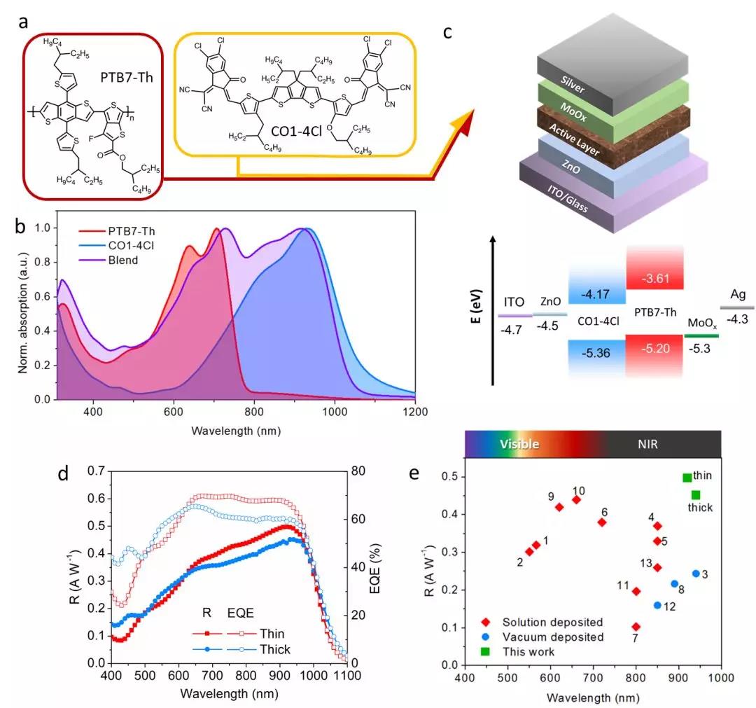
The active layer material of the photodetector is critical to light responsiveness. The authors selected the organic polymer semiconductor PTB7-Th as the electron donor material, and combined a new narrow bandgap (-1.2 eV) non-fullerene small molecule as the electron acceptor material CO1-4Cl (Fig. 1a). The active layer of the heterojunction of the photodetector body gives it a good visible-near-infrared absorption spectrum (Fig. 1b). The energy levels of PTB7-Th and CO1-4Cl form a typical interdigitated interstitial relationship (Fig. 1c), which facilitates efficient charge separation of photogenerated excitons at the donor-acceptor interface. For the photodetectors with two active layer thicknesses, an external quantum conversion efficiency of 60%-70% can be obtained in the red-light-induced near-infrared region at a small reverse bias of 0.1 V (Fig. 1d). The working photodetector has better near-infrared light responsiveness than the organic pn junction-based non-gain photodetector that has been reported in other literatures (Fig. 1e).
Figure 1. (a) chemical structure of the organic semiconductor of the photodetector active layer material and (b) absorption spectrum of the film; (c) schematic diagram of the basic structure and band structure of the photodetector; (d) photoresponsiveness of the photodetector And external quantum efficiency; (e) Comparison of the highest light responsiveness of this paper with other literature.
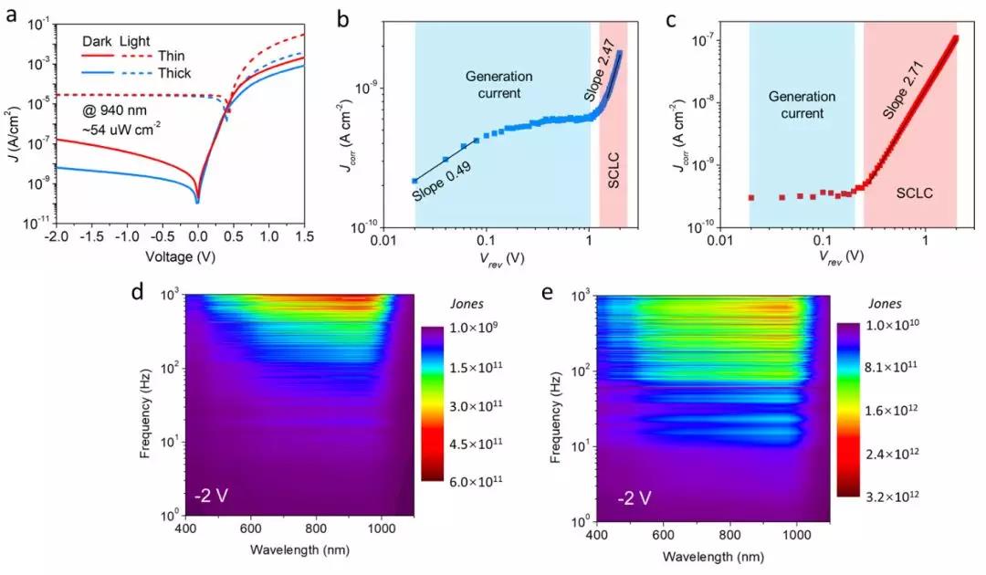
In applications with very weak signal detection, the limiting factor often comes from noise signals caused by dark currents. In order to suppress dark current and noise signals, the authors used a "thick heterojunction strategy" to achieve a low dark current of nanoamperes per square centimeter in a thick active layer device under a reverse bias of 2 volts (Fig. 2a). This low dark current is due to the effective suppression of the space charge limited current (Figure 2b-c). In combination with the high light responsiveness previously described, the ratio of 10^12 Jones can be maintained even at large reverse bias (2 V) (Fig. 2d-e). That is, it can detect extremely weak 940 nm near-infrared light signals with power densities as low as about a few picowatts per square centimeter (less than one billionth of the AM 1.5G solar power density).
Figure 2. (a) photodetector photocurrent and dark current; (b) thick active layer and (c) thin active layer photodetector to correct current density versus reverse bias; (d) thin active layer and (e) Detection rate of junction detectors with thick active layers
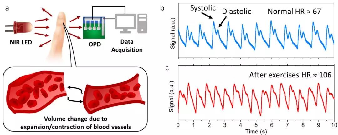
The authors further applied the prepared photodetector to the photoplethysmography method (Fig. 3a). In this method, a portion of the human body (usually a finger) is first illuminated with a beam of near-infrared light, and the transmitted/reflected near-infrared light is collected and detected by a photodetector (Fig. 3b-c). Useful clinical medical information such as heart rate, heart rate variability, etc., can be obtained due to changes in light absorption/scattering caused by relaxation and contraction of blood vessels during the cardiac cycle.
Figure 3. (a) Basic principle of photoplethysmographic pulse detection; (b) (b) normal and (c) post-exercise pulse signals detected by photoplethysmography based on photodetector
background
Near-infrared light generally refers to electromagnetic waves from 750 nm to 1400 nm. It is in the red light band of visible light in the spectrum of electromagnetic radiation, and belongs to a section of infrared light of short wavelength. Although invisible and intangible, near-infrared detection plays an important role in many technologies. For example, automated control, night vision monitoring, bioluminescence imaging, infrared photography, etc. all require efficient infrared light detectors. Near-infrared photodetectors typically rely on narrow-bandgap inorganic semiconductor materials such as single crystal silicon, germanium, indium selenide/gallium. However, such devices are generally complicated in process, expensive in cost, poor in mechanical properties, and high in temperature sensitivity. In comparison, solution-processable organic semiconductors enable the production of cost-effective near-infrared photodetectors. Its soft material properties are also beneficial for its application in flexible devices for easy integration into wearable sensor systems.
At present, organic photodetectors that are optically responsive to near-infrared light still have problems of low photoelectric conversion efficiency and insufficient detection capability for extremely weak signals. For this reason, increasing the external quantum efficiency of the device and suppressing the dark current under the detection conditions are the key to further improving the performance of the organic photodetector.